

Group website:
Other company websites:
Choose website







Forensic X-Ray Systems for Medical Examiners: A Practical Guide to Postmortem Radiography


Forensic X-Ray Systems for Medical Examiners: A Practical Guide to Postmortem Radiography
How can digital X-ray imaging help medical examiner and coroner offices handle growing caseloads without turning every case into a CT examination? Explore how modern forensic radiography supports trauma documentation, foreign-object localization, identification and more efficient postmortem workflows.
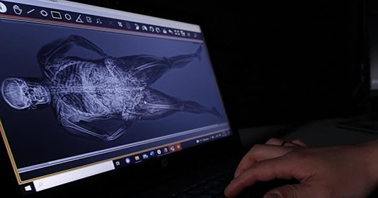
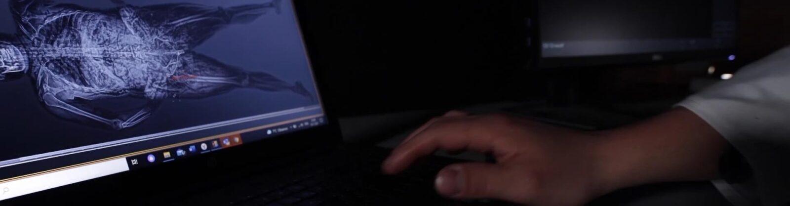
A forensic X-ray system gives medical examiners, coroners and forensic pathologists a fast way to document what is inside the body before an invasive examination begins. Unlike clinical radiography, which supports diagnosis and treatment of a living patient, postmortem radiography is designed around evidence documentation, trauma assessment, identification and medicolegal death investigation.
That distinction matters in the United States, where many medical examiner and coroner offices are being asked to handle growing caseloads with limited forensic-pathology resources. A whole-body digital X-ray examination can provide an immediate overview of fractures, projectiles, implanted devices and other internal findings before dissection, helping the forensic team decide what requires closer examination and how the case should proceed.
Modern forensic X-ray systems are therefore not intended to replace the forensic pathologist. Their value is in making the investigative workflow more informed before irreversible steps are taken.
What Is a Forensic X-Ray System?
Forensic radiography uses X-ray imaging to answer investigative rather than clinical questions. Instead of asking how an injury should be treated, the forensic team may need to know:
- Where is a projectile or metallic fragment located?
- Which bones are fractured?
- Is a fracture consistent with the reported circumstances?
- Is an orthopedic implant present that could assist identification?
- Is there a foreign object that should be documented before recovery?
- Does the case require additional imaging or a more extensive autopsy?
The underlying X-ray physics are familiar, but the equipment and workflow are different. A system intended specifically for forensic use needs whole-body coverage, appropriate table capacity, flexible positioning and digital documentation suitable for case review rather than routine clinical patient care.
Does Forensic X-Ray Replace an Autopsy?
No. Forensic X-ray should be treated as an imaging and documentation layer within a larger medicolegal investigation.
Where state law, agency policy or the circumstances of the case require a complete autopsy, a radiograph does not remove that requirement. Instead, imaging gives the pathologist additional information before dissection and leaves behind a permanent visual record of internal findings.
This is particularly valuable because an image acquired before an invasive procedure preserves the original relationship between skeletal injuries, projectiles, foreign objects and other structures. That record can later be reviewed by another pathologist, radiologist, investigator or court expert without requiring access to the remains themselves.
Forensic X-Ray vs. Postmortem CT
Digital radiography and postmortem CT are complementary technologies rather than direct substitutes.
CT provides cross-sectional and three-dimensional information and is especially valuable in complex trauma, ballistic reconstruction and cases where depth relationships are critical. Full-body X-ray provides a much faster projection-based overview and requires less infrastructure.
| Imaging Method | Primary Strength | Typical Forensic Use | Operational Consideration |
| Digital forensic X-ray | Fast whole-body projection imaging | Fractures, projectiles, foreign objects, implants and initial case assessment | Less depth information than CT |
| Postmortem CT | 3D cross-sectional anatomy | Complex trauma, ballistic reconstruction and detailed anatomical investigation | Greater acquisition, infrastructure and operating requirements |
| MRI | Advanced soft-tissue visualization | Selected specialist investigations and research | Longer examinations and greater operational complexity |
| 3D surface scanning | Detailed external documentation | Surface injuries and scene/body documentation | Does not show internal anatomy |
For many medical examiner offices, the more useful question is therefore not “X-ray or CT?” but “Which cases actually require CT?”
A rapid full-body radiograph can serve as an initial imaging layer for routine cases. Cases where three-dimensional reconstruction adds meaningful investigative information can then be referred for CT. This approach allows a facility to reserve the more resource-intensive modality for cases where its capabilities are genuinely needed.
Why Modern Forensic Imaging Uses Digital Radiography
Digital radiography, or DR, replaces film with an electronic detector that converts the X-ray signal directly into a digital image.
For forensic operations, this changes the workflow substantially. Images become available immediately after acquisition and can be reviewed, enlarged, adjusted and stored without waiting for film processing.
Digital files can also become part of a broader evidence and case-management workflow. They can be archived, reviewed remotely, incorporated into PACS environments and shared with qualified specialists when a second opinion is required.
This is particularly useful in jurisdictions covering large geographic areas where the forensic pathologist, radiologist and investigating agency may not all work in the same facility.
What Can a Postmortem X-Ray Reveal?
A whole-body forensic X-ray can reveal several classes of findings that are important during death investigation.
Skeletal Trauma
Fractures and dislocations can be documented throughout the body before dissection. In motor-vehicle collisions, falls, blunt-force trauma and other injury cases, this can help the pathologist understand the overall distribution of skeletal damage before focusing on individual anatomical regions.
Bullets and Metallic Fragments
In firearm cases, radiography can show where a projectile or fragment is located before recovery. This provides an objective record of its original position and can help guide the subsequent examination.
Medical Devices and Implants
Orthopedic hardware, prostheses and other implanted medical devices can provide useful identification information when compared with available medical records.
Unexpected Foreign Objects
Whole-body imaging can also identify objects that might otherwise remain unnoticed until the examination is underway.
A U.S. example comes from Montgomery County Forensic Services in Texas, where FOBOS XR imaging identified a firearm and loaded magazine among a deceased individual’s belongings during a case examination. The radiograph provided information that was relevant not only to the investigation but also to staff safety before further handling. See the Montgomery County FOBOS XR case study.
Where X-Ray Fits Into the Medical Examiner Workflow
A practical postmortem imaging workflow can begin before the body is extensively manipulated.
- Case intake and identification. Case information and imaging identifiers are assigned.
- Initial external examination. The forensic team documents the condition of the remains.
- Whole-body radiography. AP, lateral or other required views are acquired.
- Image review. Trauma, foreign objects, implants and other relevant findings are identified.
- Examination planning. The pathologist decides what additional imaging or invasive examination is required.
- Evidence recovery. Radiographic findings can help guide recovery of projectiles or other objects.
- Archiving. Digital images become part of the case record for later review.
The benefit is not simply speed. Imaging provides information early enough to influence how the rest of the examination is conducted.
FOBOS XR: Full-Body Forensic Radiography
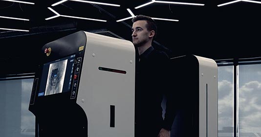
FOBOS XR is a purpose-built full-body digital radiography system for morgues, medical examiner offices, coroners and forensic centers.
The system is designed to acquire a whole-body X-ray image in less than 30 seconds and supports a table capacity of up to 340 kg / 750 lbs. AP imaging is standard, with lateral and angled imaging capabilities available for more detailed examination.
Because the platform was developed specifically for postmortem work, the table, imaging geometry and workflow are designed around deceased subjects rather than adapted from routine clinical patient positioning.
Typical applications include:
- full-body skeletal assessment;
- fracture documentation;
- projectile and fragment localization;
- medical implant identification;
- pre-autopsy examination;
- case documentation;
- research and forensic-pathology education.
When Full-Body X-Ray Can Be More Practical Than CT
CT remains the appropriate imaging modality when true three-dimensional anatomical reconstruction is necessary. However, not every case requires that capability.
A dedicated forensic X-ray system can cover a large volume of routine postmortem examinations with substantially less infrastructure than a CT suite. This is relevant for county medical examiner offices and forensic centers evaluating not only equipment purchase price but also installation, room requirements, staffing, maintenance and long-term operating cost.
The operational objective is not to make an X-ray system imitate CT. It is to use a faster and simpler modality for cases where projection radiography already provides the information needed, while preserving access to CT for the cases that benefit from it.
Three Forensic X-Ray Configurations for Different Facilities
Forensic organizations operate under very different physical and operational constraints. LINEV Systems addresses three common deployment models.
FOBOS XR — Permanent Forensic Facility
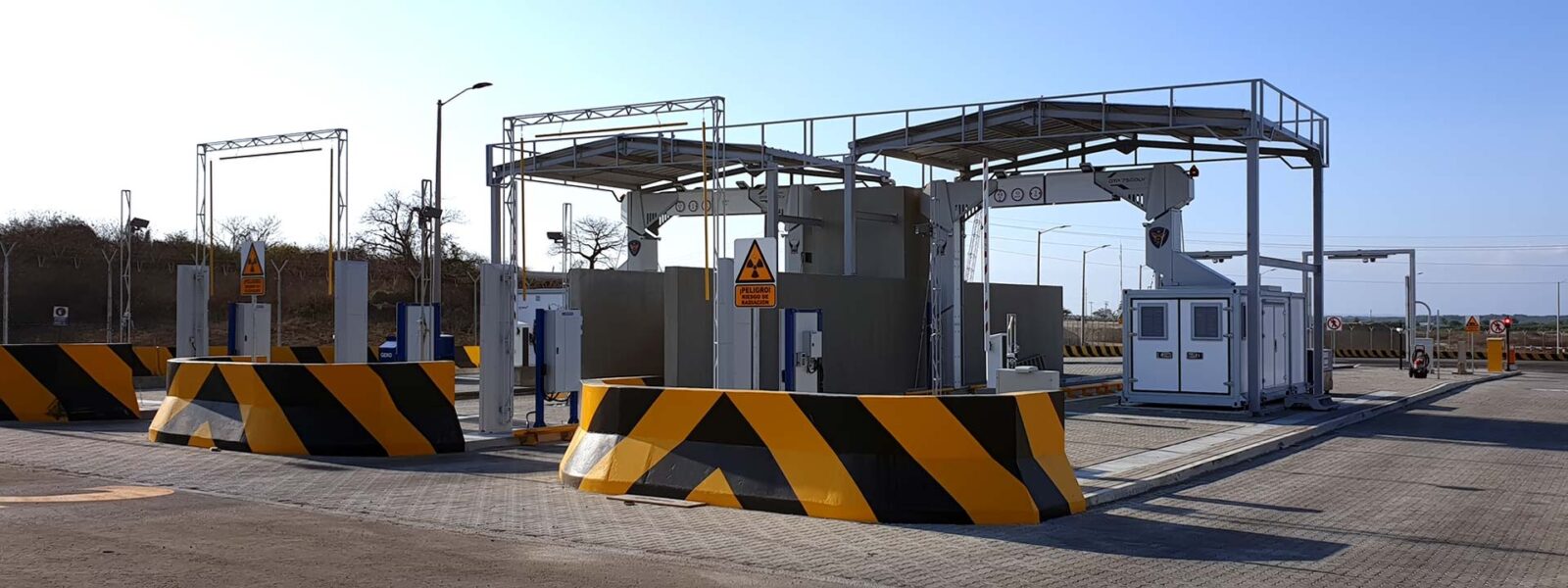
The stationary FOBOS XR configuration is intended for medical examiner offices, morgues and forensic centers with an established imaging location and ongoing caseload.
FOBOS M — Mobile Forensic Imaging
FOBOS M brings postmortem radiography to the field. A mobile configuration can support disaster victim identification, mass-fatality response and temporary forensic operations where transporting all remains to a permanent imaging facility would delay the investigation.
FOBOS XRS — Shielded Forensic Radiography
FOBOS XRS incorporates radiation shielding into the system configuration. This can be particularly relevant for facilities that do not already have a dedicated shielded X-ray room or where a major construction project would complicate deployment.
Mobile X-Ray for Mass-Fatality and DVI Operations
A mass-fatality incident changes the imaging problem completely. Instead of bringing a small number of cases into a permanent morgue, forensic teams may need to examine a large number of remains at or near a temporary processing location.
Mobile digital radiography allows imaging to become part of that field workflow. Skeletal injuries, foreign objects, implants and other identifying information can be documented before extensive handling.
Radiographic records are also relevant to disaster victim identification because ante-mortem and post-mortem imaging can contribute to identification alongside dental records, fingerprints and DNA.
For these situations, the value of FOBOS M is not only that it produces an X-ray image. It moves the imaging capability to the operational site rather than requiring every case to be transported to an established radiology department first.
PACS, Digital Evidence and Case Review
The image is only one part of the forensic workflow. How that image is stored, retrieved and associated with the case is equally important.
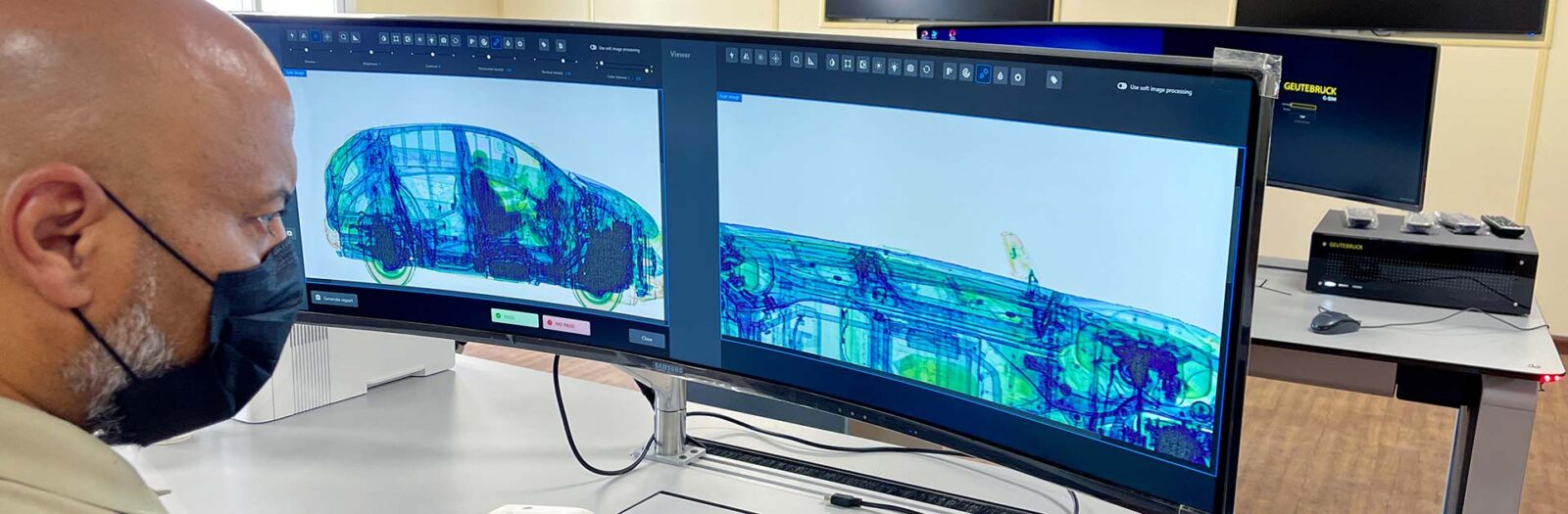
A digital forensic radiography system can support PACS-based archiving and specialist review. This allows the original image to remain preserved while qualified users apply viewing tools such as zoom, contrast and window adjustments without modifying the original acquisition data.
For procurement teams, PACS compatibility should therefore be evaluated alongside image quality and scanning speed.
How to Evaluate a Forensic X-Ray System
Medical examiner and coroner offices should begin with their operational profile rather than a single specification.
Important questions include:
- How many cases does the office process annually?
- How often is whole-body imaging required?
- What body-weight capacity is needed?
- Are AP and lateral images required routinely?
- Is there an existing shielded room?
- Does the system need to integrate with an existing PACS environment?
- Is mobile DVI or mass-fatality deployment part of the agency’s responsibility?
- What training is required for technologists and forensic staff?
- What service and parts support is available in the United States?
These factors usually determine whether a stationary FOBOS XR, shielded FOBOS XRS or mobile FOBOS M configuration best fits the agency.
FAQ: Forensic X-Ray Systems
What is a forensic X-ray system?
A forensic X-ray system is a digital radiography platform designed for postmortem examination and medicolegal death investigation. It can document skeletal injuries, projectiles, implants and other internal findings without an incision.
Do medical examiners use X-rays during autopsy?
Yes. Radiography may be used before or during a forensic examination to identify fractures, locate foreign objects and document findings that help guide the autopsy.
Can forensic X-ray replace CT?
No. CT provides three-dimensional cross-sectional information that projection radiography cannot. X-ray can, however, serve as a rapid first-line imaging method so CT is reserved for cases where its additional depth information is important.
Can X-rays locate bullets in a body?
Yes. Metallic projectiles and fragments are readily visible on radiographs, making X-ray useful for documenting their location before recovery.
Can a forensic X-ray help identify unknown remains?
Yes. Surgical hardware, orthopedic implants, healed fractures and other skeletal features can provide information that may be compared with ante-mortem records as part of a broader identification process.
Does a forensic facility need a dedicated X-ray room?
That depends on the system configuration and applicable radiation-protection requirements. Stationary systems may be installed in an appropriately designed imaging room, while shielded configurations such as FOBOS XRS are intended for facilities where integrated shielding is preferable.
What is the difference between forensic radiography and forensic radiology?
Forensic radiography refers to acquiring the images. Forensic radiology refers to interpretation of medical imaging within the legal and investigative context of a forensic case.
A Faster First Look Before the Autopsy Begins
Whole-body forensic radiography does not replace professional judgment, autopsy or advanced imaging when those methods are required. Its advantage is that it provides objective internal information early in the case.
For medical examiner and coroner offices, that means fractures can be mapped, projectiles located, implants documented and unexpected findings identified before the examination changes the original condition of the body.
Facilities evaluating a forensic imaging program can review the complete LINEV Systems FOBOS forensic X-ray range and compare stationary, mobile and shielded configurations against their caseload, infrastructure and workflow.
Sources
1. National Academies / National Institute of Justice analysis: Strengthening the U.S. Medicolegal Death Investigation System.
2. INTERPOL: Disaster Victim Identification Guide.
3. Montgomery County Forensic Services: FOBOS XR case study.

Manage Consent
Functional Always active
The technical storage or access is strictly necessary for the legitimate purpose of enabling the use of a specific service explicitly requested by the subscriber or user, or for the sole purpose of carrying out the transmission of a communication over an electronic communications network.
Preferences
The technical storage or access is necessary for the legitimate purpose of storing preferences that are not requested by the subscriber or user.
Statistics
The technical storage or access that is used exclusively for statistical purposes. The technical storage or access that is used exclusively for anonymous statistical purposes. Without a subpoena, voluntary compliance on the part of your Internet Service Provider, or additional records from a third party, information stored or retrieved for this purpose alone cannot usually be used to identify you.
Marketing
The technical storage or access is required to create user profiles to send advertising, or to track the user on a website or across several websites for similar marketing purposes.